
 New advances in dental technology have made it so that dentists and orthodontics can do more than ever before. The usage of X-rays is one of the most useful tools that these professionals have, and there are numerous types of X-rays that dentists take to help them determine the source of a patient’s dental issues. Not only do X-rays assist in determining the causes of dental issues, but they also greatly assist in the treatment planning of those issues. One of those types of X-rays is known as a Ceph X-ray.
What Is a Ceph X-ray?
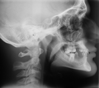
A Ceph X-ray is actually the shortened term for the technical term Cephalometric X-ray. A Ceph X-ray is used to capture a radiological image of the side of a patient’s face. This type of X-ray is different from a “full set” X-ray in that it allows the dentist or orthodontist to more clearly view just the side of the face, including the teeth, jawbone and soft tissue beyond what can be seen in a full set X-ray and also beyond what can be seen with the naked eye. This type of X-ray is also extraoral, which means that no plates or films are used in taking them. Perhaps the biggest advantage that Ceph X-rays offer over intraoral ones is that they allow the dental professional to get a view of the nasal and sinus passages as well, which aren’t displayed on full set, bitewing or other intraoral X-rays.
What Is a Ceph X-ray Used For?
Ceph X-rays are primarily used to provide orthodontic diagnosis that is used to plan treatment for an orthodontic surgery, most especially an orthognatic surgery. This type of X-ray helps the orthodontist assess progress throughout the treatment period and serves as a handy guide for reference when needed. Most importantly, it allows the orthodontist to determine whether or not the dental issue that the patient suffers from is due to skeletal reasons, dental reasons or both. The X-rays can also be used for research purposes, but in order for this to happen, they need to be clinically certified first. The most common reasons that dentists and orthodontist cite for using a Ceph X-ray is that they provide views of the following:
• The side profile of the face.
• The jaw in relation to the cheekbone.
• Information about “bad bites” or malocclusions.
• Measurement of the teeth.
• Fractures and other injuries to the teeth and jawbone.
How Are Ceph X-rays Taken?
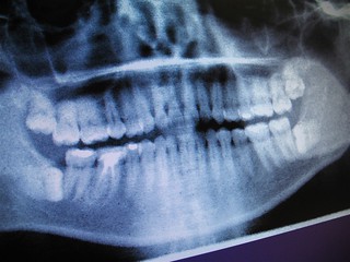 Ceph X-rays are taken with a panoramic X-ray machine that has a special Cephalometic film holder mounted underneath a mechanical arm located on the machine. Then, the X-ray film is exposed to ionizing radiation that will provide the dentist with a picture of the entire side of the oral structure. This type of X-ray is painless and merely consists of the patient placing his or her head into plastic ear rods and a chin holder, both of which work to stabilize the head and hold it into perfect position as the X-ray is conducted. It only takes a few minutes, and then the process is over with. Once the X-ray is produced, it’s important that the dental professional trace the image to help him or her get a clearer picture of the distance between each part of the oral structure. Once that’s done, then the dentist can determine whether the patient possesses an anteroposterior relationship or a vertical one. An anteroposterior relationship usually shows up after the age of seven and indicates whether or not the patient suffers from an overbite while a vertical relationship is merely based upon the proportions of the facial height and will indicate whether or not the patient suffers from an underbite. The way that the dentist can determine this is by measuring the angles at which all the proportions of the face come to as well as analyzing and interpreting other skeletal factors.
References:
Marie C. Trocmé ; A. Howard Sather ; Kai Nan An. “A biplanar cephalometric stereoradiography technique.” Retrieved on February 9, 2016, from https://mayoclinic.pure.elsevier.com/en/publications/a-biplanar-cephalometric-stereoradiography-technique(5a42a84f-f549-48e1-bf4c-9fb720de6925).html.
Devereux L1, Moles D, Cunningham SJ, McKnight M. “How important are lateral cephalometric radiographs in orthodontic treatment planning?.” Retrieved on February 9, 2016, from http://www.ncbi.nlm.nih.gov/pubmed/21300228.
Mayo Clinic
5777 E Mayo Blvd.
Phoenix, AZ 85054-4502
480-515-6296
http://www.mayoclinic.com
National Center for Biotechnology Information, U.S. National Library of Medicine
8600 Rockeville Pike
Bethesda, MD 20894
888-346-3656
http://www.ncbi.nlm.nih.gov/
Images:
https://farm3.staticflickr.com/2154/2299189555_43a6be71ce_n.jpg
https://farm1.staticflickr.com/179/465952448_c815300562_n.jpg